
Tag: Medicine

New system for detecting Parkinson’s early

Parkinson’s disease is a debilitating neurodegenerative disease, affecting everything from speech, posture and gait to digestion, sleep, impulse control and cognition. Therapies exist that alleviate some symptoms of the disease, but there is still no cure for Parkinson’s, which affects close to one million Americans and 10 million people worldwide.

A new Tel Aviv University study unveils a novel method for detecting the aggregation of the protein alpha-synuclein, a hallmark of Parkinson’s disease. With this knowledge, caregivers could introduce treatment that has the potential to significantly delay disease progression.

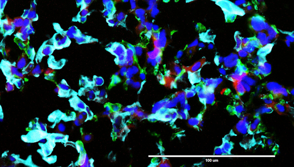
By the time a patient is diagnosed with Parkinson’s disease, 50 percent to 80 percent of the dopaminergic cells in the part of the brain called substania nigra are already dead, possibly due to development of toxicity as result of alpha-synuclein aggregation. “We have developed a new method for tracking early stages of aggregation of alpha-synuclein using super-resolution microscopy and advanced analysis,” says Prof. Uri Ashery, co-author of the study and head of TAU’s Sagol School of Neuroscience and TAU’s George S. Wise Faculty of Life Sciences. The research was published in Acta Neuropathologica on May 31.

“Together with our collaborators at Cambridge University, who developed a special mouse model for Parkinson’s disease, we were able to detect different stages of the aggregation of this protein,” Prof. Ashery explains. “We correlated the aggregation with the deteriorating loss of neuronal activity and deficits in the behavior of the mice.”

A big step towards early detection

“This is extremely important because we can now detect early stages of alpha-synuclein aggregation and monitor the effects of drugs on this aggregation,” says Dr. Dana Bar-On of the Sagol School of Neuroscience, a co-author of the study. “We hope that this research can be implemented for use in the early diagnosis of Parkinson’s in patients. We’re currently working to implement the methods in a minimally invasive manner with Parkinson’s patients.”

The researchers, in collaboration with the Max Planck Institute in Gottingen and Ludwig-Maximilians-Universität München, were able to illustrate the effect of a specific drug, anle138b, on this protein aggregation and correlated these results with the normalization of the Parkinson’s phenotype in the mice, according to Prof. Ashery. “This is a significant step forward in the world of Parkinson’s research,” he says.

The researchers are planning to expand their research to family members of Parkinson’s disease patients. “By detecting aggregates using minimally invasive methods in relatives of Parkinson’s disease patients, we can provide early detection and intervention and the opportunity to track and treat the disease before symptoms are even detected,” Prof. Ashery concludes.

Could diet and exercise help prevent dementia?

A new Tel Aviv University study published in the Journal of Alzheimer’s Disease finds that insulin resistance, caused in part by obesity and physical inactivity, is also linked to a more rapid decline in cognitive performance. According to the research, both diabetic and non-diabetic subjects with insulin resistance experienced accelerated cognitive decline in executive function and memory.

The study was led jointly by Prof. David Tanne and Prof. Uri Goldbourt and conducted by Dr. Miri Lutski, all of TAU’s Sackler School of Medicine.

“These are exciting findings because they may help to identify a group of individuals at increased risk of cognitive decline and dementia in older age,” says Prof. Tanne. “We know that insulin resistance can be prevented and treated by lifestyle changes and certain insulin-sensitizing drugs. Exercising, maintaining a balanced and healthy diet, and watching your weight will help you prevent insulin resistance and, as a result, protect your brain as you get older.”

A two-decade study

Insulin resistance is a condition in which cells fail to respond normally to the hormone insulin. The resistance prevents muscle, fat, and liver cells from easily absorbing glucose. As a result, the body requires higher levels of insulin to usher glucose into its cells. Without sufficient insulin, excess glucose builds up in the bloodstream, leading to prediabetes, diabetes, and other serious health disorders.

The scientists followed a group of nearly 500 patients with existing cardiovascular disease for more than two decades. They first assessed the patients’ baseline insulin resistance using the homeostasis model assessment (HOMA), calculated using fasting blood glucose and fasting insulin levels. Cognitive functions were assessed with a computerized battery of tests that examined memory, executive function, visual spatial processing, and attention. The follow-up assessments were conducted 15 years after the start of the study, then again five years after that.

The study found that individuals who placed in the top quarter of the HOMA index were at an increased risk for poor cognitive performance and accelerated cognitive decline compared to those in the remaining three-quarters of the HOMA index. Adjusting for established cardiovascular risk factors and potentially confounding factors did not diminish these associations.

“This study lends support for more research to test the cognitive benefits of interventions such as exercise, diet, and medications that improve insulin resistance in order to prevent dementia,” says Prof. Tanne. The team is currently studying the vascular and non-vascular mechanisms by which insulin resistance may affect cognition.

Breakthrough in Battle Against Brain Cancer

Groundbreaking research from Tel Aviv University may lead to a significant breakthrough in the battle against the deadliest type of cancer in the central nervous system, Glioblastoma. Glioblastoma is aggressive, invasive and fast growing. It is resistant to existing treatments, with patients dying within a year of the cancer’s onset. Glioblastoma is defined as a ‘cold tumor’, which means that it does not respond to immunotherapeutic attempts to activate the immune system against it.

Identified & Neutralized: Failure in Brain’s Immune System

To begin with, the researchers identified a failure in the brain’s immune system, leading to the amplification of cell division and spread of Glioblastoma cancer cells. The failure results partially from the secretion of a protein called P-Selectin (SELP), which, when bound to its receptor on the brain immune cells, alters their function so that instead of inhibiting the spread of cancer cells, they do the opposite, enabling them to proliferate and penetrate brain tissues.

At the next stage of the study, the researchers were able to inhibit the secretion of the SELP protein, thereby neutralizing the failure in the immune system, restoring its normal activity, and blocking the spread of this incurable cancer.

The international research team was led by Prof. Ronit Satchi-Fainaro, Director of the Cancer Biology Research Center and the Head of the Cancer Research and Nanomedicine Laboratory at Tel Aviv University’s Sackler Faculty of Medicine. The findings were published in the leading scientific journal Nature Communications.


Glioblastoma cancer cells

‘SELP Identification’: Why the Brain’s Immune System Doesn’t do its Job

Launching the study, the researchers wanted to understand why the cells of the brain’s immune system (called microglia) do not inhibit the cancer. Led by PhD student Eilam Yeini, they compared healthy brain tissues with glioblastoma tissues. To do this, they collaborated with neurosurgeons from the Tel Aviv Sourasky Medical Center (Ichilov) who supplied Glioblastoma tissue samples removed during surgery and also with neurosurgeons from Johns Hopkins University and the Lieber Institute in the USA, who supplied healthy brain tissues from autopsies.

“We wanted to understand why the brain’s immune system doesn’t do its job,” says Prof. Satchi-Fainaro, who won the Youdim, Bruno, Humboldt and Kadar Family Awards for Outstanding Research in 2020.  “We examined the interactions between the immune cells in the brain and the Glioblastoma cells in tumors that were recently removed from patients’ brains. To our surprise, we found that not only do the microglia cells do nothing to stop the cancer cells, they actually play a crucial and negative role by accelerating the division, spread and mobilization of glioblastoma cells.”


Since cells communicate with each other through proteins, the researchers checked what proteins are secreted when the microglia immune cells encounter the Glioblastoma cells, finding six proteins that are overexpressed. At the next stage, Prof. Satchi-Fainaro and her team blocked each of the six proteins in turn, seeking to identify and isolate the one that enables the cancer to harness the brain’s immune system to its own ends. Ultimately they discovered that a protein called SELP is responsible for disrupting the functions of the immune system and boosting Glioblastoma tumors.

“SELP is a known protein that normally helps cells travel inside the body – especially white blood cells and endothelial cells that line the interior of blood vessels,” explains Prof. Satchi-Fainaro. “The encounter between Glioblastoma cells and microglia cells causes them to express SELP in large quantities. In the study, we were able to show that the overexpressed SELP helps the cancer cells travel and penetrate the brain tissue.”

After inhibiting SELP in pre-clinical Glioblastoma models, the researchers found that the tumor cells had a slower division rate, stopped migrating and were less invasive. These results were attainedin animal models and in 3D cancer models. Single-cell RNA sequencing, in collaboration with Dr. Asaf Madi’s laboratory at the Department of Pathology at Tel Aviv University’s Faculty of Medicine, showed a decrease in the cancer cells’ malignant properties, and an activation of the immune system against the tumor when SELP was silenced and the communication between the microglia and Glioblastoma was disrupted. As a result, the cancer’s progression in the brain was hindered.


Awaiting Approval to Start Saving Lives

Prof. Satchi-Fainaro emphasizes that the new study may have lifesaving therapeutic implications. She mentions that, by sheer coincidence, a clinical trial phase 2 currently underway is attempting to inhibit SELP for another purpose altogether – treating pain associated with sickle cell anemia. Prof. Satchi-Fainaro hopes that the fact that the treatment inhibiting SELP has been proven safe in humans, will pave the way for relatively rapid approval of a clinical trial repurposing the new treatment for Glioblastoma. “Unfortunately, Glioblastoma patients need new treatments immediately. Our treatment may be the needed breakthrough in the battle against the most daunting cancer of all.”


The new study was funded by the Israel Cancer Research Fund (ICRF), the European Research Council (ERC), the Morris Kahn Foundation, the Israel Cancer Association (ICA) and The Israel Science Foundation (ISF).


The researchers thank the donors and their next of kin for the provision of brain tissue for this study.

TAU Team Reverses Early Signs of Alzheimer’s

Approximately 50 million people worldwide live with Alzheimer’s or other related forms of dementia. Alzheimer’s disease leads to memory loss and impairment in cognitive function, and is the most common cause of dementia among older adults. While certain treatments can help reduce symptoms and sometimes reduce disease progression, there is currently no way to prevent or cure Alzheimer’s.

Amid that backdrop, researchers from Tel Aviv University have developed a process for reversing the precursors of the disease, providing a promising foundation for new preventative therapies. This marks the first time that a non-drug therapy has proven effective in preventing the core biological processes that lead to the development of Alzheimer’s, providing hope that we will now be able to fight one of the greatest challenges to the Western world.

Targeting the Root of Alzheimer’s

Using hyperbaric oxygen therapy (HBOT), in which subjects breathe 100% oxygen in a special chamber of high atmospheric pressure, the researchers were able to reverse brain damages associated with the biological hallmarks of Alzheimer’s.

“By treating the root problem that causes cognitive deterioration with age, we are in fact mapping out the way to prevention,” says co-lead researcher Prof. Shai Efrati.

Often used to treat carbon monoxide poisoning and infections that starve tissues of oxygen, hyperbaric therapy, when applied in a specific way, has previously been found capable of repairing damaged brain tissue and renewing growth of blood vessels and nerve cells in the brain. Therefore, the researchers tested its potential for Alzheimer’s.

“After a series of hyperbaric treatments, elderly patients who were already suffering from memory loss showed an improvement of blood flow to the brain as well as a real improvement in cognitive performance,” said co-lead investigator Prof. Uri Ashery.

The new approach devised by the researchers unequivocally improved characteristics commonly associated with Alzheimer’s disease. Specifically, the hyperbaric treatment resulted in:

  • Improved memory in 16.5% of patients on average
  • Increased blood flow in 16%-23% of cases
  • Improved attention and concentration in 6% of patients
  • Improved information processing speed in 10.3% of all cases

A Future Without Alzheimer’s?

“Our findings provide hope that we will now be able to fight one of the greatest challenges to the Western world. According to our findings, hyperbaric therapy given at a young age is likely to prevent this severe disease entirely,” explains TAU team member Dr. Ronit Shapira.

The approach was first tested in laboratory settings followed by testing in patients over the age of 65 in stages of deteriorating mental function that often precede Alzheimer’s and dementia. The therapy included a series of 60 treatments in hyperbaric chambers over a period of 90 days.

The study is part of a comprehensive research program focused on reversing processes of aging and its accompanying ailments. The researchers note that the findings are an encouraging step toward new approaches to preventing Alzheimer’s by addressing not only the symptoms or targeting biomarkers, but the core pathology and biology responsible for the disease’s development.

The Tel Aviv University team that led the study included Prof. Shai Efrati of the Sackler Faculty of Medicine and the Sagol School of Neuroscience, Prof. Uri Ashery and  Dr. Pablo Blinder of the The George S. Wise Faculty of Life Sciences and the Sagol School of Neuroscience, and Dr. Ronit Shapira and Dr. Amir Hadanny. They are all affiliated with the Shamir Medical Center. The findings were published in the journal Aging.

Breakthrough TAU Discovery Key to Reversing ALS

A Tel Aviv University-led research team has uncovered a core mechanism that causes ALS and has succeeded in reversing its effects. While the root cause of ALS remains unknown, the discovery reveals the basic biological mechanism that leads to nerve destruction in the early stages of the incurable disease that afflicts an estimated one out of every 400 people.

To date, there is no effective treatment to prevent or halt disease progression. The average life expectancy of ALS patients is approximately three years from diagnosis. “This discovery can lead to the development of new therapies that could enable nerve cells to heal before irreversible damage occurs in the spinal cord,” said lead investigator Prof. Eran Perlson of the Sackler Faculty of Medicine and the Sagol School of Neuroscience at TAU.

New Tool for Combating the Disease

The team discovered that an abnormal buildup of a protein called TDP-43 in neuromuscular junctions, which translate brain signals into physical movements, leads to the degeneration and death of nerve cells (motor neurons). They found that this hinders the activity of mitochondria, which are critical for cells to function.

The researchers found that this process occurs during the early stages of ALS, initiating damage to motor neurons before patients develop serious symptoms. Eventually, the deterioration of nerve cells in the brain and spinal cord causes ALS patients to gradually lose voluntary muscle ability, leading to complete paralysis including the inability to breathe independently.

Reversing the Domino Effect

Using an experimental molecule (originally developed to enhance neural regeneration after injury), the team demonstrated its success in dismantling the toxic protein buildup found in ALS patients. Additionally, in lab models, the researchers showed that this approach actives the process of nerve regeneration, leading to almost complete rehabilitation from the disease.

Together with Dr. Amir Dori, director of the clinic for neuro-muscular diseases at Sheba Medical Center, and scientists from the US, UK, Germany and France, Perlson and doctoral students Topaz Altman and Ariel Ionescu conducted the study through a series of experiments. The findings were published in the peer-reviewed journal Nature Communication.